

Biopsy at 14 years of Age
Page 3
Microscopic Findings
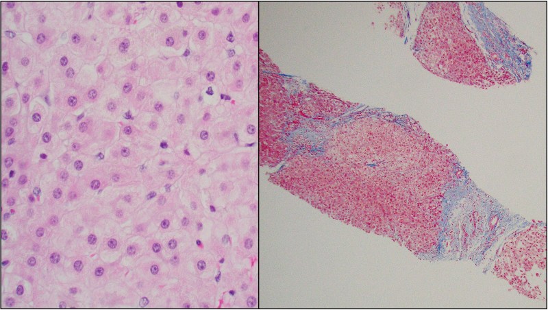
- Nonspecific reactive changes in hepatocytes
- More advanced fibrosis with bridging
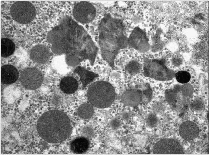
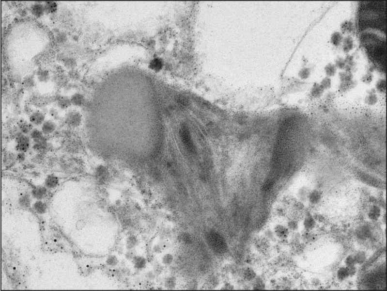
Ultrastructural Findings
References
- Warren M, Mierau G, Wartchow E, Shimada H, Yano S. Histologic and ultrastructural features in early and advanced phases of Zellweger Spectrum disorder (infantile Refsum disease). Ultrastruct Pathol. 2018 May-Jun;42(3):220-227.
- Braverman NE, D’Agostino MD, Maclean GE. Peroxisome biogenesis disorders: biological, clinical and pathophysiological perspectives. Dev Disabil Res Rev. 2013;17:187–196.
- Braverman NE, Raymond GV, Rizzo WB, et al. Peroxisome biogenesis disorders in the Zellweger spectrum: an overview of current diagnosis, clinical manifestations, and treatment guidelines. Mol Genet Metab. 2016;117:313–321.
