Exploring Pediatric Pathology

Helping Pathologists Help Kids

Search


Case 8: 15-year-old Female with Leg Pain

Go to Page 2, Home

Page 1

Go to: Clinical Presentation, Light Microscopy, Diagnosis

Clinical Presentation

Chief Complaint: 15-year-old female with leg pain

    Past Medical History: Unremarkable

    Pre-biopsy Radiology Finding:

    Well-circumscribed lytic lesion with thin septae in the distal femur

    Go to: Clinical Presentation, Light Microscopy, Diagnosis

    Histologic Findings

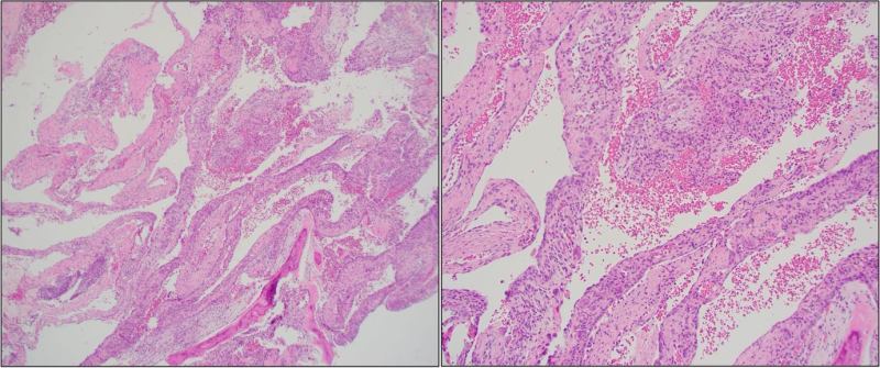
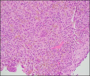
    H&E stained sections demonstrated thin fibrous septa.
    The septa comprised of fibrous stroma with spindled cells, osteoclastic giant cells, lymphohistiocytic inflammatory cells, extravasated red blood cells, and scattered hemosiderin deposits.
    Focal solid areas are seen.
    Reactive bone formation and dystrophic calcification were occasionally seen. 

    Immunohistochemical Staining

    • Not relevant

    Diagnosis?

    Go to Page 2, Home

    Pages: 1 2

    Leave a comment

    Website Built with WordPress.com.