Exploring Pediatric Pathology

Helping Pathologists Help Kids

Search


Case 6: 14 year-old Male with Hip Pain

Go to: Page 2, Home

Page 1

Go to: Clinical Presentation, Light Microscopy, Diagnosis

Clinical Presentation

Chief Complaint: 14-year-old male with hip pain

Past Medical History: Unremarkable

Pre-biopsy Radiology Finding:

Round lytic lesion in the femoral head

Go to: Clinical Presentation, Light Microscopy, Diagnosis

Histologic Findings

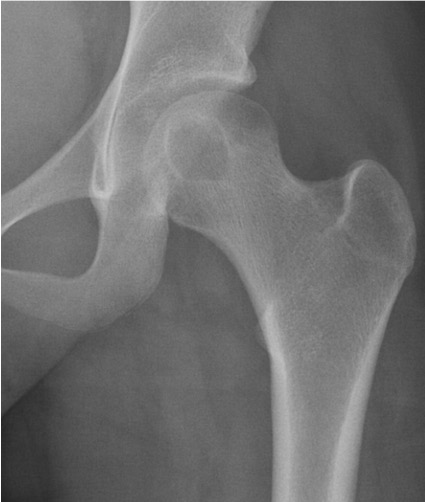
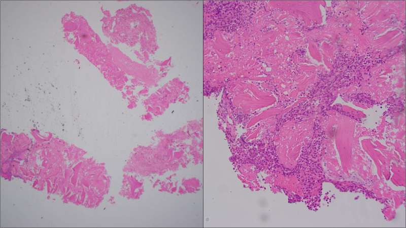
Interpretation of bone biopsies can be difficult. This biopsy contained mostly crushed bone fragments. Only a focal small area showed the lesional cells.
The lesional cells are also crushed, but they appear to have monotonous oval nuclei and abundant eosinophilic cytoplasm. No pleomorphic cells. No mitoses.

Immunohistochemical Staining

Have to rule out LCH (CD1a, Langerin: negative; S100: positive in a subset of cells (nuclear and cytoplasmic)
H3.3 K36M: Diffuse strong nuclear positivity in the lesional cells; H3.3 G34W: negative

Diagnosis?

Go to Page 2, Home

Pages: 1 2

Leave a comment

Website Built with WordPress.com.